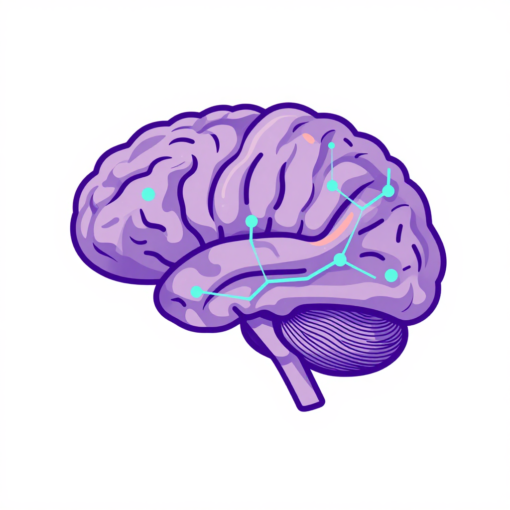
3D Brain Tissue Models
The breakthrough came quietly, buried in a Nature Neuroscience paper, but it matters enormously for anyone watching how we age. After nine years of grinding laboratory work, researchers at LMU University Hospital in Munich built a three-dimensional human brain tissue model that actually works—and works reliably enough that pharmaceutical companies could soon use it to screen thousands of drug candidates.
What makes this model remarkable isn't just that it contains neurons, astrocytes, and microglial immune cells. It's that these cells self-organize into functional mini-tissues within a single week, expressing the exact genes and proteins we'd expect to see in a living human brain. And when researchers triggered Alzheimer's pathology—amyloid beta aggregates—the model responded with active microglial clearance, just as a healthy brain would during normal cognitive aging.
"This is precisely the area in which his team has now made major progress," Dominik Paquet, Professor of Neurobiology at LMU's Institute for Stroke and Dementia Research, told Neuroscience News. "With potentially far-reaching consequences for the development of new drugs against Alzheimer's disease."
Why Three-Dimensional Brain Tissue Matters for Understanding Normal Cognitive Aging
Traditional two-dimensional cell cultures grow flat on plastic dishes. They're useful for basic biology, sure. But they completely miss the spatial complexity, multi-cell communication, and structural interactions that define how a human brain actually functions.
When you're studying normal cognitive aging—or trying to understand what goes wrong when that process derails into Alzheimer's disease—the difference between 2D and 3D isn't academic. It's everything.
The new 3D brain tissue model brings neurons, astrocytes, and microglia together in a spatial framework that mirrors how these cells interact in vivo. Neurons form extensions and connect through functional synapses. Astrocytes supply nutrients to their neighbors. Microglial cells, the brain's resident immune system, actively monitor the environment, clearing dead cells and foreign matter before they accumulate.
"It took us nine years," Paquet explained, "to develop our new, three-dimensional model of human brain tissue before it worked at all necessary levels."
The "all necessary levels" phrase is doing heavy lifting here. It means the researchers didn't just grow cells. They grew a system where every primary gene and protein associated with Alzheimer's pathology and normal cell-to-cell signaling is fully active. That's a high bar. Most in vitro models fail to capture even a fraction of that complexity.
The Tri-Culture System: How Stem Cells Self-Organize Into Working Brain Tissue
Here's where the science gets genuinely fascinating. The starting material is human stem cells, specifically, induced pluripotent stem cells (iPSCs). These cells can be converted into various brain cell types. The trick is getting them to become the right types, in the right proportions, and to organize themselves into functional structures.
The LMU team developed a proprietary differentiation cocktail, a very specific recipe of substances applied to the stem cells in a special nutrient solution. The differentiated cells then connect and adhere to each other, self-organizing into functional mini-tissue spheroids within one week.
These spheroids are approximately the size of half a pinhead. Small enough to be unremarkable, apparently. But they're doing extraordinary work.
The three cell types in the tri-culture system each play distinct, critical roles:
Neurons build functional synaptic connections. These are the communication highways of the brain, and in the 3D model, they form actual networks, not just isolated cells firing randomly.
Astrocytes support cell metabolism and structure. They're the glue, the fuel supply, and the maintenance crew all rolled into one. Without them, neurons don't survive long, and they certainly don't function properly.
Microglia are the immune cells responsible for monitoring brain health and clearing toxic protein aggregates. In normal cognitive aging, microglia shift their transcriptional landscape in response to disease signals. The 3D model captures this dynamic behavior, which previous models simply couldn't replicate.
"Almost everything just like in a real human brain," Paquet said. "We also tested whether all genes and proteins that are important for the study of Alzheimer's disease are active in our tissue model. And that was indeed the case."
Inducing and Clearing Alzheimer's Pathology: Proof the Model Works
The real test of any Alzheimer's research model isn't whether it looks like brain tissue. It's whether it behaves like diseased brain tissue, and whether interventions actually work.
The LMU team successfully triggered the formation of amyloid beta aggregates within the tissue spheroids. These are the characteristic protein clumps that define Alzheimer's disease pathology. Then, and this is the critical part, they treated the diseased 3BTMs with anti-amyloid immunotherapy, the same class of drugs now approved for clinical use.
The result? The deposits cleared. The microglia, which play a central role in Alzheimer's disease, were demonstrably active in the clearance process. And crucially, the treatment largely reversed disease signatures in the glial cells.
"This demonstrates that our model isn't just a static snapshot of brain tissue," Paquet noted. "It's a dynamic system that responds to therapeutic intervention in ways that mirror what we see in human patients."
The implications are enormous. For the first time, researchers have a reproducible platform that can model both the physiological state of normal cognitive aging and the pathological shifts that occur in Alzheimer's disease. This means drug developers can test candidates in a realistic human cellular environment before moving to animal studies or clinical trials. For readers interested in the broader landscape of Alzheimer's risk and protective factors, see Building Resilience Against Alzheimer's Disease: Risk Factors and Protective Strategies.
Robotic Automation: Scaling Brain Tissue Models for Industrial Drug Testing
The laboratory is currently adapting the platform for automated robotic manufacturing. The goal is to produce hundreds or even thousands of tissue spheroids with identical disease symptoms.
Why does scale matter? Because industrial drug testing requires consistency. You can't compare drug effectiveness if every batch of test tissue looks different, behaves differently, or has different cell compositions. Reproducibility isn't a nice-to-have, it's a prerequisite for meaningful data.
"Our system could help accelerate the development of new drugs," Paquet said. "With this goal in mind, his team is currently working on automating and scaling the manufacture of the tissue models using robots."
This robotic manufacturing pipeline addresses one of the most persistent problems in neuroscience research: the inability to generate large quantities of standardized, human-relevant tissue for testing. Animal models have their uses, but they often fail to predict human responses. Human cell cultures have improved, but they've lacked the structural and functional complexity of actual brain tissue. This 3D model bridges that gap.
For pharmaceutical companies, the ability to test thousands of potential drug candidates rapidly in a human system could shave years off development timelines and reduce the staggering costs associated with late-stage clinical trial failures.
What This Means for Cognitive Health in Older Age
Let's step back for a moment and consider what this research means for anyone concerned about cognitive health as they age.
We now understand more about what normal cognitive aging looks like at the molecular and cellular level. We know which genes and proteins are actively involved in maintaining healthy brain function. We understand how microglia shift their behavior in response to pathology. And now, thanks to this 3D model, we have a tool that can replicate all of these processes in a controlled, reproducible environment.
This isn't just about treating Alzheimer's disease. It's about understanding the mechanisms of normal cognitive aging so thoroughly that we can intervene before pathology takes hold. The model can be engineered to study both the physiological and pathological states of human brain tissue, opening doors to preventive strategies we simply couldn't develop before. For practical guidance on maintaining cognitive health in older age, explore How Midlife Vascular Health Buys You 13 Dementia-Free Years and Understanding Cognitive Aging: Why Blood Cells Enter the Brain.
The research was published in Nature Neuroscience (DOI: 10.1038/s43856-026-01767-4) by a team including Julien Klimmt, Carolina Cardoso Gonçalves, Dominik Paquet, and many others. Their work demonstrates that stem-cell-based in vitro models, when done correctly, can offer genuine insights into human brain cell functions and interactions.
The nine-year development timeline tells us something important about scientific progress: breakthroughs don't happen overnight. They require patience, persistence, and a willingness to keep trying even when the path forward isn't clear. For researchers, patients, and families affected by Alzheimer's disease, that persistence has finally paid off.
The 3D brain tissue model isn't a cure. It's not even a drug. But it is a tool, a powerful, reproducible, human-relevant tool, that could accelerate the discovery of treatments for one of the most devastating diseases facing aging populations worldwide. And in the fight against cognitive decline, tools like this are worth their weight in gold.