The Retina Has Backup Wiring Nobody Can Trigger
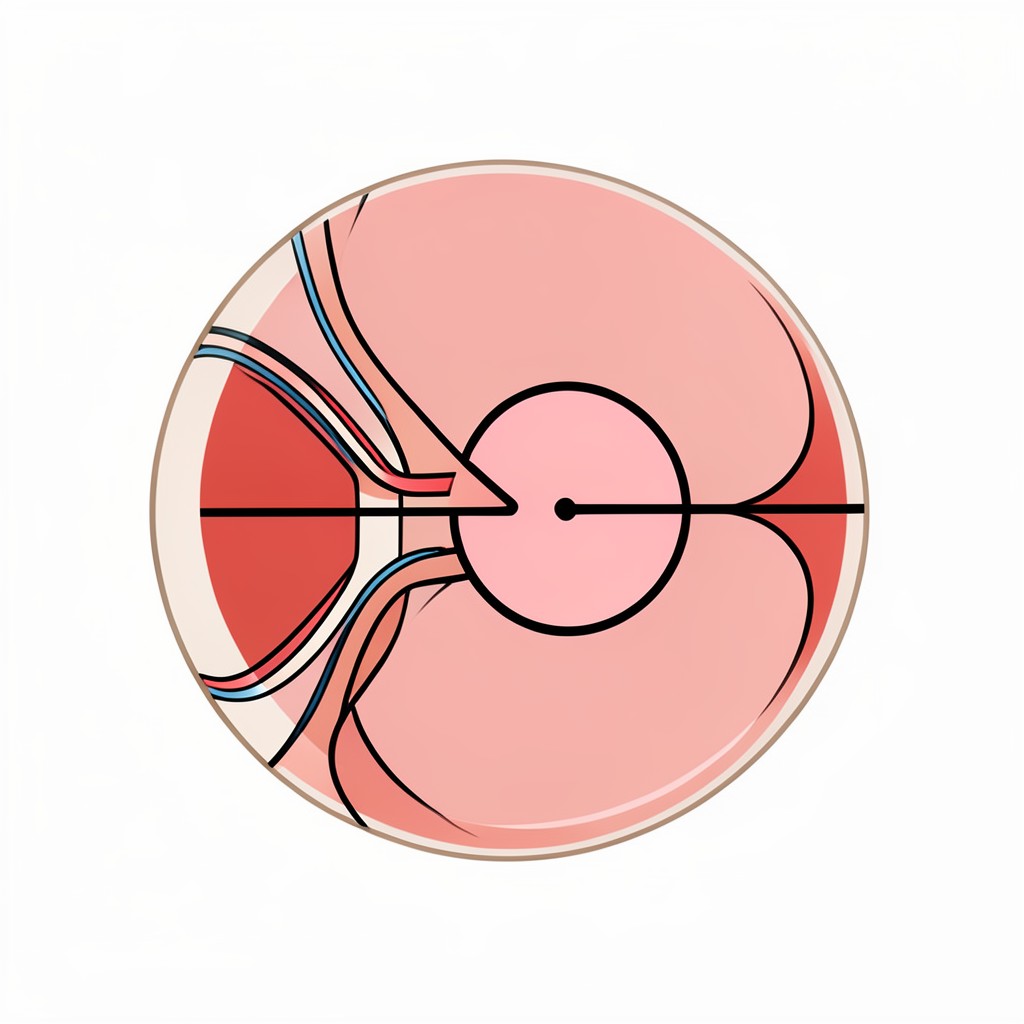
Retinitis pigmentosa robs people of sight by killing photoreceptors — the rod and cone cells that convert photons into electrical impulses. But here is the part most people miss: deeper in the retina, a second layer of neurons called retinal ganglion cells often survives intact. They are still wired to the brain. They just stopped receiving any signal upstream.
Neuroengineers have spent over a decade trying to recruit those surviving cells. Gene therapy works only for specific mutations. Optogenetics needs viral vectors to install light-sensitive proteins into the cells themselves. Electronic implants are bulky, require surgery, and degrade. Every approach hits a bottleneck because each one is fighting biology with biology.
What if you skipped the biology entirely? That is the bet a team led by Associate Professor Menglin Chen at Aarhus University just validated in a paper published in Nature Biomedical Engineering.
A Nanoparticle Drug Delivery System That Works Like a Tiny Solar Panel
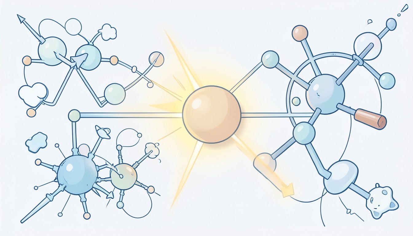
The researchers engineered hollow-sphere nanoparticles made from graphitic carbon nitride — a semiconductor that happens to be unusually good at absorbing visible light. At roughly 300 nanometers in diameter, each particle is about the size of a large virus. The architecture borrows from chloroplasts: a hollow shell that maximizes surface area for photon capture.
Once injected near retinal ganglion cells, these particles settle into position and begin spontaneously internalizing into nearby cells with what the paper describes as excellent cytocompatibility. No viral vector. No genetic modification. The particles are inert until light hits them.
And when light hits, they generate local physical and chemical reactions — both photoelectrochemical and photothermal — that depolarize the adjacent neurons. The cells fire. The signal propagates up the optic nerve to the visual cortex. The brain sees something.
That is not metaphorical. The team measured it.
Visual Cortex Activation and Behavioral Proof in Blind Mice
In mice engineered with advanced retinitis pigmentosa, animals that are functionally blind, the team injected the hollow graphitic carbon nitride nanoparticles directly into the vitreous adjacent to the ganglion cell layer. Then they shined light into the eyes.
Electrophysiology recordings from the visual cortex showed measurable, reproducible responses. The brain was receiving input. On top of that, the mice displayed observable behavioral changes consistent with light detection, not just isolated neural firing.
That combination matters. Plenty of studies produce a cortical signal in a paralyzed or anesthetized preparation. Here the animal moved differently. It reacted. The signal was functional.
Porcine Tissue Confirms the Mechanism Scales
Mouse retinas are thin. Human retinas are thicker and more layered, which makes researchers rightly nervous about whether a mechanism that works in a small animal survives translation.
The team addressed this by applying the nanoparticles to porcine retinal tissue ex vivo. Pig eyes are closer to human anatomy in scale and structure. Under light-emitting diode stimulation, the particles directly activated retinal ganglion cell activity in the porcine tissue, confirming the mechanism is not a rodent-specific quirk.
This is still not a clinical trial. But ex vivo porcine validation is a meaningful checkpoint between "works in mice" and "injectable in a person."
Why the Mutation-Agnostic Angle Changes the Commercial Math
Gene therapy for inherited retinal disease is a precision instrument. Each approved therapy targets one or a handful of mutations. The addressable population per product is narrow. Development costs are enormous. The math is brutal.
This approach sidesteps that entirely. It does not care which gene went wrong. The nanoparticles exploit whatever surviving neurons are still present, regardless of the upstream genetic cause. Retinitis pigmentosa alone spans dozens of mutations. A single intervention that works across all of them would be a fundamentally different regulatory and commercial proposition from a single-gene therapy.
That does not make it easy. It still needs safety data, delivery optimization, and long-term stability studies. The team explicitly lists these as upcoming priorities before any human application. But the strategic position is genuinely different from mutation-matched gene correction.
Beyond the Eye: Cardiomyocytes and Subcellular Control
The same nanoparticles were tested on cardiac cells. Using a focusing laser, the team achieved intracellular optical stimulation with subcellular resolution, triggering calcium-transient release in multiple cell types. At the tissue level, light-emitting diodes synchronized the beating of cardiomyocyte clusters.
The authors frame the platform as a general-purpose tool for photo-modulation spanning subcellular, intercellular, and tissue-level scales. The retina is the most clinically urgent application, but the underlying capability, light-driven cellular activation without genetic engineering, opens doors well beyond ophthalmology.
For a broader look at how nanoparticles navigate biological environments to reach target cells, see our earlier piece on the protein corona as a targeting interface.
What Still Needs to Happen
Long-term ocular biocompatibility. The particles need to demonstrate they do not cause chronic inflammation, oxidative stress, or immune rejection in a living eye over months and years. The cytocompatibility data so far is encouraging but short-term.
Delivery optimization. The current injection method places particles near ganglion cells, but uniform coverage across a human-sized retina is a separate engineering problem. How many particles are needed per square millimeter of retinal surface? Do they aggregate? Do they clear too fast?
Stability under repeated illumination. Semiconductors degrade. Graphitic carbon nitride is stable in the lab, but inside an eye bathed in ions, proteins, and oxidative byproducts, behavior changes.
None of these are dealbreakers. None of them require new physics. They require the grinding, unglamorous translational work that separates a Nature paper from an FDA filing. The team is working on it.
The Bottom Line
This is not a bionic eye. It is not a chip. It is a powder made of carbon and nitrogen that you inject and then illuminate. When light hits it, nearby neurons fire. In blind mice, that firing reaches the visual cortex. In pig retinas, the mechanism holds up.
For a field that has spent years on increasingly complex biological interventions, the elegance here is refreshing. Sometimes the right answer is not better biology. It is better physics.
We have covered other nanoparticle approaches to retinal disease, including lipid-replacement strategies that slow degeneration, but this work stands apart because it replaces function rather than merely slowing loss. Watching where it goes from here is worth paying attention to.